Knee Cartilage Repair Options
Cartilage treatments that are considered focal have several treatment options. General degeneration of the overall cartilage, such as later stage osteoarthritis, has different treatment paradigms. In this section, we consider focal cartilage treatment options. In all cases, consult with your doctor to determine which treatment option is right for you.
Non-Surgical Knee Cartilage Repair Options
Once a cartilage injury has been diagnosed by your doctor, typically through MRI or visual examination (via arthroscopy), palliative (or non-surgical) options may include non-steroidal anti-inflammatories, like aspirin or ibuprofen, rest and elevation, and physical therapy.
Surgical Knee Cartilage Repair Options
There are many surgical treatment approaches to cartilage repair recognized in the United States. The most common of these are the marrow stimulating techniques. More recently, the use of autologous or allograft tissues are being explored. Autograft or autologous tissue refers to your own tissue. The term allograft refers to tissue from a donor (not self).
Cartilage Repair Options
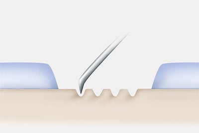
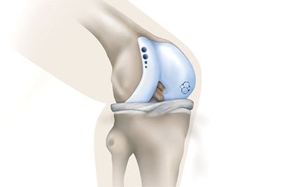
In marrow stimulating techniques, the surgeon removes damaged cartilage and pierces the bone beneath with a drill, high speed burr or awl to cause the bone to bleed. The bone marrow, blood and cells create a “clot” in the cartilage defect. Stem cells within the clot fill in the defect with fibrocartilage.
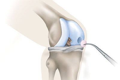
In these techniques, the surgeon will either use a plug or multiple plugs of cartilage from an area of the joint that does not bear weight to replace damaged cartilage. In some cases, tissue from a donor will be used to replace damaged cartilage. The allograft tissue is tested for disease prior to use in a patient.
Autologous cartilage implants involve harvesting and expanding the patient’s own cartilage producing cells, called chondrocytes, and then implanting them back into the patient in a two step procedure. There are two different techniques to achieve autologous cartilage implantation.
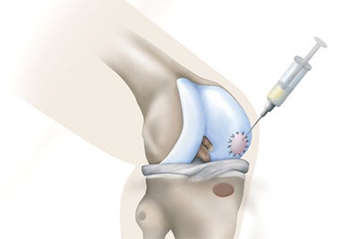
Classical ACI procedures require the harvest of the patient’s own cartilage producing cells. The cartilage samples are taken back to the manufacturing facility where the cells are removed and provided with the right environment to multiply. When enough cells have been made, they are ready to go back to the surgical center. During the second surgery a piece of thick membrane is harvested from the top of the tibia to be used as a cover to hold the chondrocytes in place. The membrane is positioned and sealed in place and the new autologous chondroycytes are injected beneath to create a “blister.”
All surgical interventions have associated risks. Please consult with you doctor to discuss risks and benefits.
